Tuesday, September 10, 2013

EpiLASIK, ICL or LASIK?

In recent years, LASIK has become one of the commonest operations performed. Its popularity and success are understandable, given the good outcomes experienced by the majority of patients. Still, there are potential side effects with any procedure, and LASIK also has its limitations especially for those with very high spectacle degrees. 2 commonly mentioned alternatives are epiLASIK and ICL (implantable collamer lens), and in Singapore these 2 procedures have been marketed quite heavily as well. Some of their proponents would have you consider undergoing these ops rather than LASIK, and in their reading material sometimes emphasise the potential side effects of LASIK. So, what's the deal with these alternatives and are they really to be preferred over LASIK?

There are pros and cons with each procedure. With epiLASIK, the advantage (which is also a disadvantage, as you will see later) is that no corneal flap is created, so theoretically the eye is stronger and perhaps at somewhat lower risk for a complication called corneal ectasia.

Having said that, the vast majority of cases that we do are LASIK rather than epiLASIK. Visual recovery following LASIK is much faster than after epiLASIK. In comparison, vision the second day after LASIK is already pretty good (usually in the region of 6/6 and 6/7.5), but for epiLASIK, to reach a comparable level of vision you might need to wait several weeks. We also take precautions not to go below 250 microns of residual stromal thickness and this avoids ectasia in normal LASIK patients (the risk being in the region of 1 in 5000 cases or so).

The problem with epiLASIK/LASEK/PRK procedures is that the corneal epithelium is removed (see below). Healing of this to provide a smooth surface will take from several weeks up to a couple of months or so. Because the surface is ablated, there is a chance of corneal scarring, but this is much less nowadays since we use mitomycin C at the end of epiLASIK/LASEK/PRK. 
EpiLASIK blade cleaves corneal epithelium from the stroma

I have grouped epiLASIK/LASEK/PRK together since they are essentially the same procedure. The difference is in how the epithelium is removed. In epiLASIK, a special blunt blade (tissue separator/microkeratome) cleaves the epithelium from the stroma. There is a risk that the blade may cut into the corneal stroma sometimes, resulting in an uneven surface, and because of this risk we don't do epiLASIK in our clinic. LASEK uses alcohol to loosen the epithelium, and this is our method of choice if we decide on surface treatment for a particular patient. The LASEK we do is 'epithelium-off', or 'alcohol assisted PRK' (see below in italics). Traditional PRK used a toothbrush type scrubber and we think the alcohol is gentler to the eye. In our clinic we currently reserve LASEK for patients with corneas that are too thin for conventional LASIK. The number of patients who have this done is probably less than 5%. The others all have LASIK with a 'bladeless' technique ie. using a femtosecond laser to create the flap and then the excimer laser to reshape the cornea.

A little bit of history: Why, might you ask, did people invent different ways to remove the epithelium when it could very easily be rubbed off with any instrument at hand? Well, it turns out that once people realised how long an eye took to recover from PRK, they tried to keep the thin epithelial cell layer intact and replace it at the end of the operation rather than throwing it away. The 2 ways of separating the epithelium while keeping it intact were 1) using alcohol to loosen the epithelium (LASEK) and 2) using a special blunt blade (epiLASIK). It turns out that once the epithelium is peeled away, whether by alcohol or the blunt blade, it dies. If this dead layer of cells is replaced, it slows healing down even more! Therefore, nowadays, usually even when LASEK or epiLASIK is done the epithelium is thrown away rather than replaced. So that makes it identical to PRK, doesn't it? Why spend money on an epiLASIK blunt blade when you can simply peel away the epithelium, perhaps with alcohol to loosen it first? There just doesn't seem to be a role for epiLASIK...and furthermore when you use a blade, even a blunt one, you run the risk of cutting into the stroma when you don't want to...
The ICL


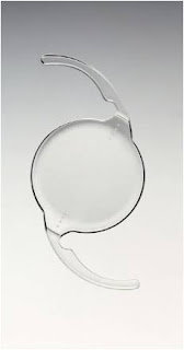
The ICL (above) is similarly different, with different pros and cons. When LASIK is done for patients with very high spectacle degrees or thin corneas, the area of cornea treated (optical zone) is smaller, so that the cornea does not get too thin. Unfortunately, when the area treated is small, the risk of halos in the dark and poorer quality vision also rises. The good thing about the ICL is that halos, even if present, are usually very mild even when the spectacle power corrected is high. It is also true that after ICL, the eye does not get as dry as after LASIK.
The Artemis ultrasound biomicroscope

However, potential intraocular (inside the eye) complications exist with the ICL, of which cataracts and glaucoma are the 2 main (but rare) ones. The main thing about reducing complications with the ICL is to achieve accurate sizing, and at our clinic we do 'sulcus to sulcus' measurements for all ICL patients. What is the sulcus? This is the little nook in the eye just behind the iris (coloured part of the eye) and in front of the original crystalline lens where the ICL will sit. If the ICL is too big for this space in a particular eye, it will be 'squashed', bend forwards, and narrow the drainage angle of the eye (increasing the risk of glaucoma). If the ICL is too small for this space, it will sink backwards and touch the crystalline lens, thus causing a cataract. Although there is no perfect way to measure this sulcus diameter down to the last micron, we have found the Artemis VHF ultrasound biomicroscope (above) to be very accurate and allows us to choose an appropriate size ICL. The figure below shows a measurement made by the Artemis.

I would say that for my patients with lower spectacle powers, LASIK provides excellent clarity of vision including night vision (and without the risk of any intraocular complications). I have performed ICL surgery for a number of patients with excellent results, but I think bearing in mind the potential seriousness of problems with the ICL my preference is still an extraocular (outside the eye)  procedure like LASIK in cases where I judge that the outcome will be good. Extraocular means zero chance of causing cataract or glaucoma.

Actually, I think the ICL is wonderful, but I prefer to use it for patients with very high powers or thin corneas, where I feel that the risks of intraocular problems are outweighed by the better visual quality that can be obtained with the ICL in such cases compared with LASIK.

Although a lens is implanted into the eye in both ICL as well as cataract surgery, an ICL op is quite different from a cataract op, not least because when an ICL is implanted the patient's original crystalline lens/cataract is left alone while during a cataract op the crystalline lens/cataract is removed and is replaced by an intraocular lens implant (IOL). This has implications because there are some doctors out there who perform 'clear lens extractions' to correct high spectacle degrees. If a 'clear lens extraction' (which is like a cataract operation) is done in a younger person less than 45 years old or so, then they will immediately lose the ability to focus for near ie develop immediately the maximum amount of presbyopia/LaoHua. ICL surgery will not affect the development of presbyopia, nor can it cure this problem. ICL surgeons can offer monovision with the ICL, just like with LASIK or contact lenses.

On the other hand, if somebody has a cataract then he/she should have a cataract op, not an ICL op. The ICL surgery doesn't do anything about a cataract, which would continue to blur the vision. 

Overall I would say, go for LASIK if your cornea is thick enough because the theoretical advantages of epiLASIK probably do not bear out clinically for the majority of patients. ICL surgery is a great option for those who would otherwise be at risk for visual or other side effects from LASIK.

Thursday, September 5, 2013

Eye doctor trivia

In Ophthalmology, as in many other fields, "innovation" is always regarded as a golden word and there are countless doctors/researchers at any time looking for better ways to treat a given eye condition. True innovation requires a number of qualities on the part of the "innovator", and perhaps it's as good a time as any to re-visit the stories behind some of the greatest inventions of the twentieth century.

He came from a family of doctors and clergy, and once was a lapchild of Florence Nightingale.

His father was an ophthalmologist, and following a distinguished stint in medical school received the FRCS (Fellowship of the Royal College of Surgeons) at the tender age of 25. Even around this time, before formal training in eye surgery, he had begun discussing the possibility of replacing crystalline lenses after cataract surgery with his father and close associates.

In 1940, Flight Lieutenant Gordon Cleaver was returning from a sortie in his Spitfire. In his haste to get into the plane that morning, he had forgotten his flight goggles, so when a bullet smashed through the canopy of his fighter, bits of perspex plastic from the canopy were blown straight into his eyes. In the end, he underwent 18 operations on his eyes and face, many of which were performed by this doctor.

It used to be said (something to the effect of) 'Thou shalt not have a foreign body in the eye'. And yet, it was quite clear in this particular patient that some of the remaining plastic pieces in the eye were not causing any problems even years after the original injury.

So this young doctor spoke with some spectacle manufacturers and eventually a small perspex disc similar in shape to the original crystalline lens was produced. This was implanted into the eye of a patient on 29 November 1949. It wasn't long before he felt the full force of hostility (and some might say jealousy) from his peers. For several decades, the topic was not even given a section for presentation and discussion at conferences. By the 1970s, though, the unquestionable benefits of implants over thick aphakic glasses were apparent and with improving lens designs, better results were also being seen around the world.
The first intraocular lens implant


Today, it is the unusual patient who does not get a lens implant. The question has become 'why didn't this patient get an implant?' rather than the other way round. And of course, this was the story of Harold Ridley and the intraocular lens implant, which has revolutionised eye surgery in fundamental ways.


He was a bit of a maverick, but a persistent one at that.

Finding himself unable to get into medical school in the United States, he applied and got accepted into the University of Geneva Medical School in Switzerland, where for a time he was spending more time composing music than on his medical studies. But fortunately for all of us, graduate he did, and became heavily involved with research into better ways of doing cataract surgery.

In the mid 1960s, he was given US$299,000 to find a way to remove cataracts through a small incision (cut). Going through more than 40 different ways of doing so, including a meat grinder (!) type of device, he failed every single time and was close to despair. " I thought I might face defeat better if I looked better, and so I prepared," this doctor wrote in his 1985 autobiography Through My Eyes. "The haircut came first, then a shoeshine, and finally, the dentist." 

At the dentist's, he was very taken with the then also new method of removing calcified plaque with an ultrasonic cleaner. After many hundreds of modifications later, including refinements in technique from many other surgical experts, we now have almost one day visual rehabilitation, with many patients achieving better than 6/9 vision on the first day after surgery.

The first phacoemulsification machine


Of course, I'm talking here about Charles Kelman, the legendary inventor of phacoemulsification, which is the gold standard for cataract surgery nowadays.


A common thread that runs through these innovators seems to have been a long standing conviction that something was not quite right and could be done a whole lot better. Although it seems so obvious to us nowadays, it took long and winding roads before conviction was translated into innovation. But for serendipity, sometimes, nothing might have come of it.

It has been said (on Ming Thein's blog and perhaps others) that he who tries 99 times and fails is seen as a fool, while success on the 100th time is a sign of great persistence in the face of adversity. Perhaps the frustration and fear of failing just before success is as good a motivating force as any!

For all the success that we see sometimes, I think the respect we accord to these pioneers should include a great dose of not just awe for their genius and intellect, but also their willingness to risk everything (peer respect and sometimes even careers) and to persist in the face of great adversity. In fact, it seems like the greater the uproar created, the greater the invention! The ability to think laterally and to find uses for seemingly unconnected things is also priceless. And to this end, I have viewed the increased sub and sub-sub specialization of the medical specialties with some concern, since the narrower world view could discourage cross-discipline exchange of ideas and methods.

As always, it is the devil in the details, finding the right balance between craziness of an idea and potential for success, and decisions regarding the distribution of limited funds, that will determine the success or failure of future endeavours.


Saturday, August 31, 2013

Computers and the Eye-Bane, Boon, or Much Ado About Nothing?

Eye and visual problems are among the most commonly reported problems among computer workers, affecting up to 70% of such a population in some studies. The term Computer Vision Syndrome (CVS) refers to a range of eye and vision problems related to near work during computer use. It is not a specific disease, but rather, a range of symptoms related to visual demands exceeding the ability of a person to comfortably perform the task. Although associated with discomfort, there is generally no permanent harm to the eyes or vision.


Things that patients feel with computer use for prolonged periods include
1) non-specific eye discomfort (often called eye-strain)
2) transient blurred distance vision
3) transient blurred near vision
4) dry or irritated eyes
5) headache, and even
6) temporary double vision.

Why is there a problem with computer work when there may be no problems reading other things?
Working at a computer requires frequent eye movements, continuous eye focusing and alignment which involve repetitious muscular activity. For example, frequent eye movements from paper documents to the computer screen and back again occur. Viewing distances and angles for computer work are often different from those commonly used for reading and other tasks, with computer work often occurring at a distance slightly greater than the normal reading distance.

Aspects of the computer display such as resolution, contrast and glare also affect visual comfort. Often the letters on a video display screen are not as sharp as those on a printed page. The presence of reflections from the screen may also make viewing more difficult.

What factors may make the problem worse?
Uncorrected long or short-sightedness, astigmatism and ‘old-sight’ can make working at the computer more difficult. ‘Old sight’ or presbyopia refers to the natural loss of focusing ability (accommodation) with age that tends to become manifest at about 40 years of age. Some of these problems may not result in symptoms under less demanding conditions and some patients are not even aware of their presence. Patients experiencing problems should have a comprehensive eye test looking at the refractive power of their eyes.

Other possible conditions include undetected latent squints and dry eye. The prolonged concentration associated with computer work often results in a reduced blink rate, which causes dry eye because of increased evaporation of tears.

Children and computers
Today, use of computers by children is increasingly common both for education and play. Although children are more adaptable than adults in many ways, they are not as self aware and may ignore problems otherwise noticed by adults. For example, children may play an enjoyable game for hours until exhaustion. As a result of prolonged eye effort, many of the problems seen in adults may eventually be seen in children as well such as focusing problems and even dry eyes. Of concern also is the possibility of prolonged near work contributing to the progression of short-sightedness in children.

Children are also smaller than adults, and many computer work stations may not fit them well. Awkward postures may result in arm, neck or back discomfort.

When children use the computer, parents may want to introduce strict limits on the amount of time they are continuously on the computer. It is also a good idea to have the child’s vision checked some time in early primary school to exclude any refractive error that can contribute to eye strain. The work station should also be checked, with attention given to raising the height of the seat so the child can work in a more comfortable position.

Tips for managing and avoiding computer vision syndrome
Owing to the fact that computer vision syndrome refers to a range of complaints associated with computer use, its management is multifaceted.

People who work on a video display terminal and easily feel tired or uncomfortable should have a vision test to check for refractive errors. Even if glasses are already being worn, they may not be the correct power for the particular distance of the computer monitor. This is especially important for the presbyopic patient. A computer is often placed at an ‘intermediate’ distance, in between a normal reading distance and the far distance. For some presbyopes, glasses for intermediate distance in addition to the normal bifocals may therefore be necessary. Occasionally a latent squint is detected and if necessary the situation may be improved with special prism glasses.


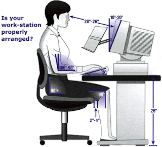
For comfortable use, the computer display should be suitably positioned. Ideally, the top of the screen is placed just below the viewer’s eye level, and tilted slightly upwards. This allows a comfortable view in slight downgaze which may help with near focusing. Environmental lighting issues are also important. When working on a computer, people should avoid facing an unshaded window, because the much brighter background would make it difficult to view the display screen. On the other hand, sitting with the back to the window with bright light shining on the screen can cause reflections. Use shades or curtains to control the amount of light entering the room.

Eye dryness often exacerbates the tired feeling many patients experience. Apart from reduced blinking, the typical office is air conditioned with very low humidity. Using artificial tears helps greatly, as does the slightly lower computer screen since the upper eyelids are lower in downgaze and the eye is less exposed to the environment.

Finally, it is a good idea to take short breaks from working at the computer. During these breaks looking out a window or closing the eyes for a few minutes rests the ciliary muscles of the eye responsible for focusing. Blinking or closing the eyes replenishes the tear film. Fortunately, despite the multitude of symptoms associated with computer use, relatively simple steps can alleviate most of the problems and the problems are generally temporary in nature.

Monday, August 26, 2013

Presbyopia treatments: Love them or hate them...multifocal lens implants for cataract patients

The last frontier with regard to presbyopia correction really has to be lens replacement surgery a.k.a. cataract surgery with lens implantation. 'New lenses for old...' to paraphrase something from Aladdin :) Here we are talking about dealing with the problem at its source.

As covered in my post about presbyopia, the underlying problem is the inability of the aging lens to focus, which boils down to its increasing stiffness with age.

For those people young enough not to know how it feels like, the next few lines might give you an idea. Basically, it creeps up on you. You thought you could always do it, you know, things like holding reading material up close to your nose, cutting your nails up close, and for the ladies-doing the mascara and eyeliner to perfection. Then one fine day you realise, it doesn't work up close anymore...it's just too hazy or you get a headache trying to focus it up close! For those in an older age group, even things like the food in front of you are not so clear anymore...

So it is easy to understand why people go to such great lengths to find a way to improve their near vision. Anyway, I digress. If the lens is the problem, let's replace the lens then!

Cataract surgery is pretty much routine nowadays with great outcomes, and a variety of lens implants are available to replace the hazy lens. If we want to provide a patient with both clear distant as well as clear, near vision without glasses after cataract surgery, there are 2 main options: monovision and multifocal lenses. Monovision refers to using a lens to make one eye have clear distance vision, and the other eye using a lens implant that makes it mildly shortsighted. That way, with one eye seeing far and one eye seeing near, both far as well as near objects are in focus. One downside is that since both eyes are not exactly seeing clearly together at a particular distance, 3-D depth perception is somewhat affected and some patients with monovision will get a pair of glasses for certain tasks such as driving.

For each eye to see well in the distance and near, multifocal lenses are a reasonable option. If both eyes are implanted with the same lens, then depth perception is not affected. One thing which needs to cleared up right from the beginning is that these lenses are not the same as progressive or bifocal spectacle lenses-ie with these lens implants, the patient does not look up to see in the distance and down to see near. Instead, the lens splits the incoming light into two focus points all the time, so there is always one in-focus image and one out-of-focus image.

The original multifocal lenses were zonal refractive lenses like the Array, which has been redesigned and renamed as the ReZoom (above). Interesting names... Zonal refractive lenses have alternating ring shaped zones, which focus light alternately for distant objects and near objects. The problem with these lenses is that they are probably the most likely of multifocal lens to cause troublesome halos and visual disturbances. They are also not that good at providing good close up vision.
Different parts of an AMO Tecnis multifocal lens


The other class of multifocal lenses are the diffractive lenses. These lenses use diffraction as a way to split the incoming light into 2 or more focus distances. Diffractive lenses have a series of sharp edged rings cut into the lens surface, and when light rays hit these edges, they will tend to spread out and form 2 different focus distances. By varying the height of these rings, the focus distance can be altered and some of these lenses have 3 focus points.

Another type of multifocal lens uses a near segment, and looks like a bifocal spectacle lens. I was quite interested in this lens but became less so when I realised it was only available as a plate haptic lens.

There is actually a 3rd option available for patients who want good near and distance vision after cataract surgery. These are the 'accommodating' lenses (which are not multifocal), and in one way or another can change their shape or the position of the lens optic in the eye. I put the word 'accommodating' in quotes, because it is unlikely that in the long run they provide much accommodation as the capsule in which they sit scars up and becomes stiffer. Even in the early period after surgery, some patients do not have as good near vision as they would like.

The option I prefer for my patients at this time is the diffractive multifocal lens. These very predictably give good near vision. And the diffractive multifocal lens I favour at this time? The Tecnis multifocal and Tecnis multifocal toric. 

(NB I do not receive any compensation from any of the lens implant manufacturers)

BUT, and this is a big but, I do not implant many multifocal lenses by any means. Most of my patients do well with monovision if they prefer not to have to use reading glasses. Clearly, there are several downsides with multifocal lenses, even if you ignore their cost.

Multifocal lenses degrade vision to some extent. Because there is always one in-focus and one out-of-focus image in the eye, the patient generally notices halos or ghosting of images to some extent. And because not all light from an object is in focus at any one time, contrast is not as good as with a normal monofocal lens. In the 2 pictures below, I've tried to give a simulation of the halos around lights caused by multifocal lenses. The first picture shows a small light source (called a 'muscle light'), and in the picture below that, I have overlayed a drawing from one of my multifocal lens patients of what he sees when I show him the muscle light. This patient has a Tecnis Toric Multifocal and has 6/4.5 unaided distance vision as well as N5 unaided near vision and is very happy. He tells me he sees 2 definite light rings around the main light, and then much fainter rings outside that. But regardless, he was expecting this, and he feels that it is a small price to pay for being able to read up close without glasses again.

Halos caused by a diffractive multifocal lens


Due to the fact that images are innately degraded by the multifocal nature of the lenses, I make absolutely sure that there is nothing else that can degrade the vision further. ie the patient cannot have any other eye disease like glaucoma or macular degeneration, and I will use the toric version of the multifocal lens as necessary to minimise post operative astigmatism as much as possible. All patients also get a pre-operative corneal topography, so that I can screen out the patients with irregular corneas (which are fairly common in the older age group).

Then I usually wait for a patient to request such a lens. And I make sure I tell them about the possible halos, and reduced contrast. Generally I will also use these lenses for patients who have denser cataracts, and who are long-sighted rather than short-sighted before surgery. My approach is extremely conservative, but I do this because the side effects are very real and patients sometimes have unrealistic expectations about what is achievable.

Why the Tecnis lenses, you might ask?

As I covered in an earlier post, I prefer lenses with flexible C loops. I also prefer the diffractive over the refractive designs as there tends to be fewer visual disturbances from halos. And that really leaves me with 2 options: The Tecnis and the Restor. The Achilles heel of the Acrysof Restor really is the problem with glistenings. This is what David Apple said:

'In summary, four clinical issues occur in eyes with glistenings. Most commonly there are subjective
complaints of poor vision in spite of a satisfactory, even normal Snellen visual acuity.

Secondly, subjective poor quality vision develops in patients who also have decreased Snellen visual acuity and/or contrast sensitivity, 

Thirdly, there is decrease or loss of the lenses special function. In such cases there is impairment of the lens’s ability to provide the ‘‘premium’’ result intended. In other words, glistenings can cause not only visual disturbances, but also may impede the designated function of a given lens. In such cases the lens may not only be affected with visual degradation, but also may be unable to provide multifocality. 

Finally, there is iatrogenic decrease in vision secondary to the additional surgical trauma of the explantation/exchange procedure.'

Apple et al. Modern Cataract Surgery: Unfinished Business and Unanswered Questions. SURVEY OF OPHTHALMOLOGY VOLUME 56 SUPPLEMENT 1 NOVEMBER–DECEMBER 2011.

David Apple was a very respected eye pathologist who specialized in lens implants. He maintained a collection of thousands of these lenses, which had been taken out from patients' eyes when they developed problems. As such, he was considered one of the foremost experts on lens implant problems. Sadly, he passed away in 2011.

Finally, multifocal lenses can result in very happy patients...or very unhappy patients. It all boils down to making sure that the patient knows what to expect, and whether they feel that the good near vision outweighs the side effects. Of course, surgery has to be as perfect as possible too...and that is up to the surgeon.

So, that's that with presbyopia options in this day and age. If there is anything I have not covered that you would like to know more about, please let me know. Incidentally I noticed that in the 'Mind Your Body' section of The Straits Times last week, there was another article about presbyopia treatments. This time it was about the Raindrop corneal inlay that I covered in my last post, and apparently there is a private practice eye centre in Singapore that is conducting a trial on it. Hmmm, having so many different inlays (the Icolens was featured in this same section of The Straits Times in January this year only) reminds me of the early days of cataract implants, when there were myriad different designs as well. If there was one design which worked really well, you wouldn't need so many different types...food for thought...

Anybody with comments about these presbyopia treatments?

Update (25 November 2014)
A new generation of lenses is just about coming online, which address presbyopia but with a different twist. Where previous lenses would split light into two focus distances, these newer lenses are called 'extended depth of field' or 'EDOF' lenses and avoid some of the problems with traditional multifocal lenses. Typically, EDOF lenses do not cause significant halos, and provide a longer continuous range of focus distances without any 'gap' in between, like intermediate distance. The first of these is the Symfony lens from AMO, but I hear HOYA has come out with one, I see a patent from Alcon about an EDOF lens, and even from Staar, which are testing an EDOF ICL!

Saturday, August 17, 2013

Presbyopia treatments: So, you want to be a spring chicken again?? (Part 1)


To follow on from the last post, so if your lens has now matured nicely in its eyeball and is no longer focusing as well as it used to, what can you do?

As with most things in life, it depends...(unfortunately, 'it depends', because there is no one size fits all, cure-all solution and all methods currently involve compromises in one fashion or other)

The simplest, time proven method is the spectacle. Spectacles are great-besides being a fashion statement, people like me gain an air of authority at work and they are also eminently adjustable and flexible. Your eye power changed by 50 degrees? No problemo, your friendly neighbourhood optometrist just needs to change the spectacle lens and Voila! everything is nice and clear again.

But...spectacles fog up, they can collect droplets of rain/sweat/etc, and if they are only used for reading they are often lost or they are not handy just when you needed them. Progressives give you a narrower field of vision and require slight adjustments of head position to work well. With contact lenses, monovision can be used (one eye corrected for far, the other corrected for near),or multifocal contact lenses can be worn to reduce the need for near reading glasses, but they are not perfect too and also there is the hassle of wearing them.

So, many clever people have thought up ideas to get around this problem while avoiding the need to wear glasses or contact lenses. Primarily the procedures can be divided into corneal operations and lens operations.

In terms of corneal operations, the most recently promoted procedures (fads? time will tell...) are corneal inlays, which are tiny pieces of special plastic implanted inside the cornea* itself.

*This is the transparent part covering the front of the eyeball.

The first of these inlays that comes to mind is the Kamra, which had been undergoing trials in Singapore some years ago. This is a very thin circular membrane with a central opening that is like a very tiny doughnut, and acts like a pinhole to sharpen images which would normally be out of focus.


The black polyvinyl pyrrolidone disc is 5 microns (0.005mm) thick and has a central 1.6mm diameter hole. Surrounding this hole is a rim which is perforated with thousands of microscopic openings that allow nutrients to pass through. This disc is implanted under a LASIK type corneal flap or into a pocket dissected within the cornea.

Most of us with some degree of long/shortsightedness or astigmatism know that if we squint or close our eyelids partially, vision can get a little sharper. This is because doing so creates a kind of ‘pinhole’ effect. Light rays entering dead centre of the cornea and eye do not have to be accurately focused to create a relatively sharp image. Similarly photographers know that if you step down the aperture to eg f11, you can increase the depth of field of the picture so that more things in the fore and backgrounds appear in focus. In fact, published articles report that many patients experienced better near vision after having had this implant.

However, photographers also know that if you step down the aperture excessively, image quality suffers. This is a result of diffraction, a physical phenomenon where light spreads out from a small point source. The smaller the light source, the greater it spreads out (in relative terms) and the blurrer an image gets. Among other factors, this is one source of the halos that patients with the Kamra inlay experience.

There are other sources of dissatisfaction as well. Excessive dryness from a thick flap, and the increased perception of floaters and other media opacities in the visual axis. Some patients developed refractive shifts (developed increasing long or shortsightedness) with the implant. It is not surprising that where such surgery is being done, there will be anecdotes of patients wanting the inlay to be removed-definitely not something either the surgeon or patient wants done after very expensive surgery! You can find a comprehensive summary from the UK National Institute of Clinical Excellence here http://guidance.nice.org.uk/IPG455/DraftGuidance and a pdf is downloadable from here http://www.nice.org.uk/nicemedia/live/13701/62162/62162.pdf

The Icolens
This is a small, 3mm diameter disc shaped implant with a 0.15mm channel in its centre to allow for the diffusion of nutrients within the cornea. The periphery of this implant has extra reading power to help with near focusing, while the centre of the implant has no power to allow for good distance vision. It is also placed under a LASIK type flap like the Kamra inlay above.


While this sounds like it could work in theory, in practice it is not so simple. Devices like this are dependent on pupil size, and if a patient’s pupil size happens to be bigger than usual then the effect is less than expected or the patient may experience significant halos from light scattering at the edge of the implant/multifocality. There is also some concern that the central channel could get blocked up in the long term from deposits that were seen to develop with other, earlier types of corneal  implants.

The Raindrop implant
This implant is so called because it looks like a droplet when implanted in the cornea (like the 2 other inlays above, also under a LASIK type corneal flap). It works by increasing the curvature of the central 2mm of the cornea, with the increased curvature helping the eye to focus for near. However, in a similar way to the Icolens, the patient's pupil size will affect the usefulness of the device and there is a very real risk of halos and distortions due to the multifocality induced by the implant. The increased central curvature on the cornea also makes that eye more shortsighted, and you can actually get this effect from plain monovision in the first place!

The only positive thing about any of these corneal inlays is that they can be removed and the procedure reversed. But personally if there was even a 2 or 3 percent chance that a patient would not like an implant and request its removal, I don't think it's worth the effort and financial cost involved, plus the blurred vision and side effects that necessitate reversal of the procedure.

PresbyLASIK
PresbyLASIK is basically LASIK, but in this case using the excimer laser to sculpt the cornea into a shape to give the effect of either the Icolens (centre-far presbyLASIK) or the Raindrop implant (centre-near presbyLASIK). Suffice to say this has not really caught on either, due to a high chance of poor quality vision/halos as above, and also the high chance of not getting the desired effect of good near vision. If anything, presbyLASIK gives less predictable outcomes than corneal inlays, and that is because the body tends to smooth out the newly sculpted shape, leading to a regression of effect.

If all of the above doesn't sound all that promising...it's mainly because it's somewhat difficult and unnatural to try and treat a lens problem on the cornea. Creating a multifocal cornea will also inevitably lead to lowered visual quality due to the aberrations (Imagine trying to take photographs with a multifocal camera lens...especially one where the effect varies as you change the aperture settings...)

It's probably too early to write off the above technologies at this time, but still it concerns me how they are being presented to the public sometimes. Goodness knows there have been so many dead ends in the last decade or so. (Remember Conductive Keratoplasty? or Intracor?)

In the second part of this series I will be giving my take on something just a little more promising-lens implants for improving near vision.

**If any reader has had corneal inlay surgery done and would like to share their experience, I would be most grateful if you could add your comments below!


Tuesday, August 13, 2013

Presbyopia: A sign you’re not a spring chicken anymore…..

Imagine you had a camera and it stopped focusing. It might take clear pictures in the distance, but up close everything becomes a hazy mess. Well, back goes the camera to the service centre or perhaps it's time to get the next iteration of your favourite DSC or DSLR (or autofocus lens...)

Spoilt camera lens

If only we could do the same with our eyes, for this is exactly what happens when the big 4-0 arrives. The lens in our eyes becomes stiffer, and this prevents it from focusing well for both far and near. In short, the eye becomes a fixed focus camera.

In actual fact, the process is a gradual one, and the lens loses its elasticity from as young as the twenties. However, it is in the early 40s when the loss of elasticity reaches such a level that focusing at reading distances becomes a pain. By the time one reaches the mid-fifties to sixties, there is virtually no focusing ability left.

The proper term for this is ‘Presbyopia’, but people may call it ‘old sight’, ‘longsightedness’, Lao Hua in Chinese, or Rabun Tua in Malay.

How it affects different people
How the condition affects you depends on what your existing spectacle power is, and what you normally use to obtain clear vision.

Let’s talk about the simplest situation, that of people who see well in the distance and do not need to wear glasses. (This includes people who have had refractive surgery like LASIK). These people continue to see well in the distance, but find that they have to hold reading material further and further away to keep it in focus. Eventually, it just becomes too tiring to read at arms length and also having to squint forcefully causes significant brow ache. That’s when reading glasses become one’s indispensable companion.

The second situation is of people who are shortsighted, ie they need glasses to see clearly in the distance. Without glasses, these people’s eyes are like cameras with extension tubes fitted. They lose infinity focus but on the other hand can focus at macro distances. Therefore, if they take off their glasses, they can see remarkably well for near even when they reach the presbyopic age group. If they wear glasses or contact lenses for distance, though, they are no different from the people in the ‘normal’ group, ie their eyes have had their extension tubes removed and with the glasses everything nearby becomes blurry too. So for looking at near things, shortsighted people either remove their glasses, or they may get glasses with a reduced distance correction.

The final situation is of people who have real longsightedness. To give a camera analogy, these people’s eyes are like cameras where the lens has been mounted too close to the film or sensor plane. To make an object at any distance clear, some degree of focusing effort needs to be put in. Even for an object at infinity, the lens needs to be focused for closer than infinity for clarity. Because these people have to put in more effort at focusing, they will notice the problem with reading earlier than others. At some point, when they are unable to focus even for distant objects, everything becomes blurry. Distant objects are blurred, and near objects even more so. Many of these people end up wearing bifocal or progressive glasses, because they always need something to clear their vision, whether for far or near objects.

Common myths
I’m shortsighted, so ‘longsightedness’ after the age of 40 will cancel out my shortsightedness
Since shortsightedness is a focusing error related to the length of the eyeball, while ‘old sight’ is a problem related to the focusing ability of the lens, they cannot cancel each other out. A shortsighted person will remain shortsighted no matter how old he or she gets. However, they have the option once presbyopic, of seeing things close up by taking off their glasses/contact lenses.

Once I start to wear reading glasses, my presbyopia/old sight/lao hua/rabun tua will get much worse very quickly. So I’m not going to wear them!
Without wearing reading glasses, many older people manage to get by, by squinting, by reading in very bright light, and by holding things as far away as physically possible. It takes a lot of effort. Once they have the reading glasses, everything gets clearer without having to put in all this effort. They may forget that they used to have to strain very hard to see, so that now, without the extra effort and without the reading glasses everything appears blurry. They may blame it on the glasses, when it was actually due to their overcompensating in the past! In actual fact, the condition slowly progresses, with or without reading glasses. Therefore, I advise my patients to wear the reading glasses if they have to and if they feel easily tired otherwise.

(PS: If the camera analogies don't quite ring a bell, feel free to clarify with me. Unfortunately I'm going through a 'gearhead' phase...)

Tuesday, August 6, 2013

An overview of cataract surgery lens implants

Cataract surgery involves removing the cloudy crystalline lens of the eye and replacing it with a perfectly clear one. Nowadays, these lenses are usually made of flexible clear plastic materials which can be folded and injected through very small incisions of 1.8-2.75mm in width. Most people are not aware that there are various brands competing in the ophthalmic market, and let the doctor make the decision as to which lens to implant in their eye. All well and good, but have you ever wondered what the factors are, that are taken into consideration when selecting a lens? Were you curious to know why your doctor chose the particular lens that was implanted in your eye?

Let's take a step back, and list the important properties of a lens that is meant to stay in the eye for the rest of a person's life.

1) It must stay transparent
2) It must be optically optimal (in harmony with corneal optics)
3) It must have a low association with posterior capsule opacification
4) It must resist the healing/contraction forces of the capsular membrane in which it sits
5) It must not cause any reaction or inflammation
Simple enough?

Let's take point 1. Current materials in use such as silicone and acrylic polymers do stay transparent for many years. In the past some hydrophilic acrylic materials became opaque after developing calcium deposits in the lens optic and had to be exchanged. Among the current lenses, the concern is increasingly with the development of so called 'glistenings' in hydrophobic acrylic lenses. These are tiny water vacuoles that develop in the lens material and have been shown to cause increased forward light scatter, which can reduce contrast. (Notice the minute white dots in the picture below) Although any hydrophobic acrylic material can have this problem, it has been reported especially in lenses made by Alcon, and these include all Alcon Acrysof lenses that are currently being used including the SN60WF (IQ lens), SN6ATT (toric lens), SN6AD1-3 (multifocal lens) and SND1TT/SV25TT (multifocal toric lens).

To be fair, not all Acrysof lenses get such significant glistenings, but it is disturbing that firstly, we cannot predict which particular Alcon lens will get a more severe problem with glistenings, and secondly, among those who develop the problem the glistenings tend to get worse with time.
glistenings in an intraocular lens
Vacuoles/glistenings in a diffractive multifocal lens

Miyata A. Water accumulation in polymers [Japanese]. IOL and RS.2007;21:59-62

The only FDA approved and certified glistening free lens currently is the Envista lens by Bausch and Lomb, which is also made of a hydrophobic acrylic material. Almost all other lenses have some degree of glistening formation after some time in the eye, but none to the same degree as what is seen with the Alcon Acrysof lenses.

What about point 2? For the photographers out there, the term 'aspheric' would ring some bells. It turns out that on average, our cornea has +0.27 microns of positive spherical aberration (at 6mm diameter), so if a lens implant has -0.27 microns of negative spherical aberration, the two would cancel out nicely leaving no spherical aberration and improved contrast sensitivity. This has been borne out in some studies, and the effect is more noticeable in dim light when the pupils are more widely dilated.

The lens implants currently in use today with such a degree of negative spherical aberration belong to the AMO Tecnis range of
lenses. Should everybody switch over to these lenses then? Well,
remember that here we are talking about an average figure, so patients with lower degrees of corneal spherical aberration, or those whose lenses might be expected to centre poorly due to pre-existing factors like weak zonules should have an aberration free lens like the Envista lens instead. The problem is that an off centre lens with any built in aberration (like negative spherical aberration in the Tecnis lens) will cause even more aberrations and blurry vision.

Most lens implants in use today are meant to be implanted in the 'capsular bag'. This is like the 'skin' of the cataract that is left behind, and it is in this thin membranous capsule that the new lens sits, right in the same position as the original cataractous lens. Sometimes the original lens cells grow and form a layer behind the new lens implant, blurring vision again. Although this can be very simply treated with a 5 minute YAG laser posterior capsulotomy, it's much better to avoid it in the first place. To this end, most lenses nowadays have a 90 degree sharp peripheral edge, so that when the capsule shrink wraps itself around the lens implant there is a 90 degree bend in the capsule which stops the migrating cells dead in their tracks.

The Tecnis Toric Lens Implant
A lens should also sit in the capsule and conform to the size of the capsule as much as possible. It is known that capsular diameters vary among different people and can be larger in people who are highly shortsighted. Flexible loop haptics (like in the lens shown above) allow the lens to be made with haptics of slightly larger diameter, since their flexibility allows them to be compressed for smaller capsules. Plate haptics (like in the lens shown below), however, are rigid and do not allow this kind of ability to adjust for different capsular sizes. They will unduly stretch smaller capsules, and if they are put into larger capsules they either decentre or may move around inside the capsular bag. Woe also to the capsule that contracts and fibroses with time-they will compress and vault a plate haptic lens, causing a refractive shift, usually in a hyperopic direction.

The last point is important but has usually been taken care of in very preliminary studies, and currently lens materials are very biocompatible. Silicone is currently less commonly used, not because it causes reactions or inflammation, but because if a patient has retinal surgery and injection of silicone oil, this oil will stick to the silicone lens andspoil it.

So, to cut a long story short, which lens would I want in my own eye? (or my dad or mum's eye?)

I would have a look at the 6mm corneal spherical aberration first. If it is highly positive, or there is a large amount of corneal astigmatism, the Tecnis lens would be ideal. If the spherical aberration was low, with little corneal astigmatism, I would go for the Envista lens and be guaranteed of no glistenings. Notice that these lenses are both flexible modified C loop designs. I would avoid plate haptic designs or those with rigid 3 or 4 haptic designs. **Would I have a multifocal lens in my own eye? I think not. Presbyopia is not a nice thing to have, but for me personally I think it would be worse to have to put up with constant halos and poor contrast which is only correctable with another surgery to replace the lens!

**Update 16th February 2015
I found myself having to clarify this last part, which I wrote in 2013, because lately I find that my patients who have had the Symfony lens are turning out to be very happy indeed. The Symfony lens is not a conventional multifocal like the Restor or Tecnis multifocal. Instead, it is called an extended depth of focus lens, where the zone of focus is increased to provide good distance and near vision. The best part of it is that there are very few or no side effects like halos. It may not give the sharpest vision when viewing things very close up, but for many people, the benefits of no side effects far outweighs this slight blurring when looking at very near things. So I think this could be my new lens of choice...

Thursday, August 1, 2013

Things you should know about cataract

Cataract is a clouding of the original lens of the eye. It is a problem that becomes more common as a person becomes older. In some studies, >90% of people over 70 years old have some degree of cataract. Cataracts can develop earlier in some people if they smoke, have diabetes, or have very high degrees of shortsightedness (greater than 1000 degrees).

Cataracts usually slowly become denser with time, and there is no medicine or eyedrop which can cure it. 

Fortunately, cataract surgery is now very advanced and refined. It is day case surgery which usually takes 30 minutes or less to complete. During surgery, the cloudy lens (cataract) is removed and a new, perfectly clear lens implant is inserted into the eye.
It used to be said a long time ago that a cataract must be ‘mature’ or ‘ripe’ (right) before it could be operated on. Nowadays, that is not true. A cataract can be operated once it affects a patient’s vision such that they have difficulties in their daily life.

As such, a pilot or someone who drives may be affected even when the cataract is fairly mild, or on the other hand a sedentary person with no specific visual requirements could tolerate a denser cataract.

When should surgery be done?

Once the cataract affects a patient’s vision and daily life, and the vision cannot be improved with glasses, surgery can be considered.
Due to advances in cataract surgery, vision can be very sharp after the operation and any existing spectacle power (long or shortsightedness, or astigmatism) can be reduced to allow for good distance vision without glasses. The spectacle power after surgery is in fact customizable, so if a patient desires good reading vision without glasses, he or she could also be intentionally left mildly shortsighted.

Multifocal lens implants do in fact exist and can provide good distance as well as near vision in the operated eye. However, by splitting the light into 2 focus distances, they reduce contrast as compared to normal monofocal lenses and also give rise to halos and other visual disturbances especially in the dark. These visual disturbances are permanent and cannot be corrected with glasses. As such, many patients who want to reduce their dependence on reading glasses after cataract surgery would do better with monovision using normal monofocal lenses.

Monovision involves using lenses that make one eye focus for distance, and the other mildly shortsighted to focus for near. In this way, the 2 eyes working together enable good distance as well as near vision. Patients with monovision still occasionally need glasses, such as when driving at night or when reading fine print for long periods of time. However, they do not experience the reduced contrast or halos that patients with multifocal lenses experience.

Is there such a thing as ‘laser cataract surgery’? For many years now, cataract surgery has been performed using ultrasound to break up the cataract, so that it could be removed through a very small incision of 2-3mm. This is the current gold standard.


In the last few years, a special laser called a femtosecond laser (above) has been created to perform a few steps of the cataract surgery. It is not possible to do the entire surgery with this laser, and it only helps the surgeon to perform several parts of the surgery. At this time, using the laser requires more time than standard surgery, costs more, may cause extra bruising around the eye, and has not been shown to have any better outcomes than the standard method of phacoemulsification using ultrasound. We have not found this laser to be useful although advances in technology in the future may change this.