Wednesday, November 18, 2015

What is it like having premium IOLs that correct for near vision after cataract surgery?

Some time ago I was asked this question on the Quora website:
What is it like having premium IOL's that correct for near vision after cataract surgery?

A very relevant question, given that nowadays there are so many lens options to choose from and also different approaches to avoid reading glasses. Here is my answer:

There are several different types of IOLs that give multifocality and good near vision as well as distance vision. I will base my answer on the most commonly used lenses-the diffractive multifocal lenses like the Alcon Restor, the Tecnis Multifocal and the Zeiss trifocal lenses.
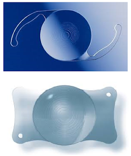
The Alcon Restor lens (above), and the AT LISA tri (below) are examples of bifocal and trifocal lens implants, respectively.


These diffractive multifocal lenses have rings etched onto either their front or back surfaces, which split/diffract incoming light rays into two foci. Some newer ones can even split the light into 3 foci. This fact gives rise to several implications.

1. Vision will be clear mainly at 2 (or sometimes 3) distances. For the older +4 Restor and the Tecnis multifocal, the near focus is at about 1 foot. So reading material has to be held quite close.

If it is held further away (say at 3 feet or so-intermediate distance), then it is not so clear anymore. 

Then things much farther away, say 15-20 feet away, start to become pretty clear again. (See the figure below-only the AT LISA tri is clear at intermediate distance)

Note: This is only the case if the surgeon has been very accurate in calculating the lens power/biometry.


Simulated clarity of images at various distances with different multifocal lens implants. The top 3 are bifocals, and the bottom one is a trifocal. Only the trifocal gives pretty good intermediate vision-but at some cost in terms of contrast when compared with the Alcon SV25T0 and AMO ZKB00. (From IOVS 2015;56:3970)

2. Because of the light splitting, there is a drop in contrast compared with a monofocal lens. Patients with multifocal lenses tend to be more sensitive to low light conditions-ie when it starts to get a little dark, they start to feel the drop in vision faster than patients with monofocal lenses. A person with a monofocal lens and reading glasses could read in dimmer conditions than a person with a multifocal lens.

3. The rings on the IOL give rings of light (halos) around light sources. This can be an issue when driving at night because of the halos around car headlamps. Having said this, many patients get used to the halos and do not notice it as much after a time.

Simulated halos around lights from a mutifocal lens

The above of course does not completely apply to the Crystalens or the Oculentis MPlus. The Crystalens is an accommodative lens, which gives somewhat unpredictable results-some patients don't get any near effect at all. 

The MPlus (available in Europe/Asia) has a wedged shaped section on the lens to focus for near. Point 1 above applies to this lens as well. However, although this lens does not give rings around lights, it can still cause 'halos' of a different shape-it is more like a smearing effect in one direction. Occasionally if a patient's pupil is small and not aligned with the lens, the patient may only have a far focus or a near focus and not both.

Last but not least, there is the Symfony lens which I covered in my last post. As mentioned, this is an 'in between' lens, ie in between a monofocal and a multifocal. It has no intermediate vision problems, and less halos than the usual multifocals, but does not give quite as good very near vision.

Currently, I offer patients a choice between a monofocal, an in between lens like the Symfony, and a full fledged trifocal like the AT LISA tri. I believe there is little to no benefit to be gained from using older bifocals such as the Restor and Tecnis Multifocal currently. Exactly which of those 3 choices gets implanted comes after a careful discussion with my patient regarding the pros and cons of each option. (So that we get big smiles all round afterwards!)

Tuesday, October 27, 2015

My experience implanting the Symfony lens this past year

What's a Symfony? Or Symphony? A symphony is actually an extended musical composition written for a variety of musical instruments, usually in an orchestra. Google Symfony and you get results pointing you to free software for a 'web application framework'.

But of course, I'll be talking about a lens implant for cataract surgery. The Symfony lens is a type of lens to allow for a greater range of clear vision after cataract surgery. Normal lens implants (monofocal) allow good vision at 1 particular distance, either near, or far, but not both. Multifocal lens implants can give good vision at 2 or 3 distances, but have not been very popular due to certain side effects like halos.


The Symfony is kind of an in-between lens-ie in between monofocal lenses and conventional multifocal lenses.

Time really flies-it's almost a year now since I started using this lens for patients. So, how's it like? In general, I would say it lives up to expectations.

In this past year, I have implanted the Symfony in different sorts of situations-including for people who already have had cataract surgery with a normal monofocal lens in the other eye.

Distance vision
With correctly done biometry, aiming for close to emmetropia (close to 0 spectacle power), the Symfony gives very good distance vision and patients were universally happy with this. 94% of my patients saw 6/6 or 6/7.5 without glasses after surgery.

The remainder had small amounts of spectacle power and in one patient whose best corrected was 6/12p, she had prior retinal detachment surgery.

What about near vision?
All Symfony lens patients could see N8 type comfortably without glasses, which is this smalland 92% could see N6 comfortably without glasses, which is this small

Although perhaps this near vision is 'less good' than the traditional bifocal or trifocal multifocal lenses, I would say this is adequate for most people most of the time...

What about halos?
Some patients, when asked, did describe mild halos around lights at night (such as car headlamps or traffic lights) but were not troubled by these halos. One patient even called it pretty!

The distinguishing feature of Symfony related halos (mentioned by 18% of my patients) seemed to be that they were quite mild/light, and patients were able to see through the halos.

Personal thoughts
There is still no perfect solution for presbyopia as yet. However, I think for myself, the Tecnis Symfony is one of the best compromises out there at the moment.

1. Minimal halos

2. Minimal drop in contrast-patients with a monofocal in one eye and a Symfony in the other are not troubled by the difference in contrast

3. Optical behaviour and patient experience mimics an early stage of presbyopia-there is no sudden fall off in intermediate vision, and vision slowly gets more blurry when things are brought closer. The effect of the Symfony has been likened to vision of somebody in their early forties.

With some of the other multifocals, there is really no equivalent in real life-the feeling could be somewhat unnatural and it could take some time to get used to.

But the Symfony is not perfect.

The chief one is as above-it is like early presbyopia. So, for people who don't mind more halos and who also don't mind a greater drop in contrast, but who would like clearer vision very close up, then a trifocal lens implant like the AT LISA tri or Finevision IOL would be good options. At the end of the day, the adage 'there is no free lunch' still holds true!


Tuesday, October 20, 2015

Learning phacoemulsification cataract surgery

Performing phacoemulsification cataract surgery is nowadays the quintessential eye operation-the one that most people think about when referring to eye doctors. To recap, this operation removes the hazy natural lens of the eye, and replaces it with a new, perfectly clear lens implant.

What's doing the surgery like?
It is a bit like playing an organ (or driving a car with manual transmission), you need both hands and both legs-how so?

Left foot:
Operating microscope (focus and zoom adjustments)
Right foot:
Phacoemulsification machine pedal (activating vacuum/aspiration and ultrasound power)


Left hand:
Second instrument for manipulating cataract
Right hand:
Phacoemulsification handpiece

So, it is a bit of a ballet, with coordination between the 2 hands and  the right leg the most important part. Like learning how to drive a car, after a while (for experienced surgeons)
1. Most of the routine parts become automatic
2. We anticipate problems that may occur
3. We consciously and subconciously set safety limits on what we do
4. We tweak our technique according to the type and hardness of cataract we are facing

Phacoemulsification is also done entirely while looking through a microscope. That takes a little time to get used to. Hand-eye coordination is particularly important, because there is much less tactile feedback when dealing with tiny things. I like to think that computer gaming in my younger days helped in this regard!

Wet lab training

A 'wet lab', you say? You mean there are wet ones and dry ones?

A wet lab in the context of surgical training involves the teaching of techniques using wet material-ie animal eyes and similar such biological tissue. We did some training for phacoemulsification on pig's eyes and also stitched pig eyelids for practice in our day.

However, animal eyes are very different from human eyes. In fact, it is usually more difficult to accomplish certain techniques with such eyes than in real life, and I was never very fond of wet labs. Still, the experience of doing things while looking through the microscope was quite useful.

Milestone: first phaco

It actually didn't take me very long before I performed my first whole cataract surgery. Besides wet labs, cataract surgery was taught (and it still is quite similar today I think) by letting the training surgeon perform some steps of the procedure at the beginning. For some trainers, this might be what are considered the easier steps first, followed by the more technically demanding ones. I started out under Mr Fearnley at Northampton doing the incision, and by the end of the year, just before Christmas 1999, I had performed my first full phacoemulsification cataract operation and lens implant under Mr Baranyovits. It was a huge milestone, finally I got the feeling that I could actually cut it as an eye surgeon!

As a trainee, there is always a sinking feeling when the supervisor says-"time for me to take over...". However, although you had to stop, it was then a great opportunity to see at first hand different techniques used to rescue the particular situation at hand, whether it was a posterior capsule rupture or zonular dialysis. The best supervisors will give a debrief and perhaps go over the surgical video when everything's done and dusted. This is a bit like reviewing the black box after a crash, the difference being that great supervisors sometimes salvage situations so well that post-operatively, the patient is none the wiser.

I went through a period when I would read anything I could find on the topic. I read Barry Seibel's 'Phacodynamics' practically cover to cover. I would visualise and imagine different techniques and scenarios in my head. I would review many surgical videos, whether they were straightforward or complicated cases. And slowly, bit by bit, doing this operation became second nature to me.

Live surgery

Every year, there are 4-5 major ophthalmic conferences where live surgery takes place. A live feed is installed from an operating theatre to the conference hall, and we get to see well known surgeons performing surgery with live commentary from a panel of experts at the conference hall. Which is not so different from listening to Thierry Henry weighing in on the Arsenal team on match day...!

I often attend these sessions, where sometimes manufacturers will showcase new surgical equipment or new lens implants. Occasionally we see new surgical techniques, but often the cases are fairly standard cataracts. Occasionally we see unplanned 'highlights' (the whole conference hall becomes deathly quiet...), and while I don't think anybody wishes for these complications, sometimes we learn more when things don't go so smoothly, rather than the other way around.

Youtube

Nowadays there are videos of everything on the internet, including cataract surgery videos! While I don't think I have learnt anything much personally from these videos, I could see how they might be useful to younger surgeons in an earlier phase of their training. It is always worthwhile to take a look and think of the things the other surgeon is doing well, and also on what could be improved.

I posted this video to share some tips to deal with difficult dense white cataracts (Note: graphic video of eye surgery):


To sum up

As in most things in life, the best practitioners of a procedure make it look simple. The best practitioners of a procedure also deal with all types of cataracts and potential complications of the procedure well. Even though nowadays cataract surgery can be done within 20 minutes or less for each eye, it takes a fair amount of time and training to get to a stage where we are both slick and safe at it. For me, it is a great privilege to have this opportunity to help so many people, and it's a very rewarding one at that!

Saturday, October 10, 2015

Medical conditions which show up in the eye

I am often asked by patients: Can you see if I have other medical problems just by looking in the eye?

Actually, yes, sometimes.

Let's start with vision. What we can see at the sides while gazing straight ahead is called our visual field. Normally we can see things about 120 degrees horizontally out in front of us, while vertically the visual field extends about 30-40 degrees.

When conditions affect the nerve of the eye, the visual field is often affected. The pattern of visual field loss can offer vital clues as to the underlying problem. For example, if the sides are affected and this loss stops abruptly at the midline, there might be a growth pressing on the nerve of the eye.

Diabetes

Diabetes is a condition which affects most if not all parts of the eye if it is not controlled well. The most well known problems have to do with retinal bleeding, which starts off with small spots of blood and fluid/cholesterol leaking into the retina, which is the thin film at the back of the eye. Later on, if it becomes more severe, new blood vessels can grow that cause more severe bleeding and scarring.

Moderate disease, with small spots of blood and some leakage of fluid with cholesterol (yellowish spots) 
Severe diabetic retinal disease with scarring of the retina
The most important factor in avoiding eye disease among diabetics is controlling the level of blood sugar. Generally, the better the blood sugar is controlled, the less the risk of geting eye disease, or of the eye disease getting worse.

High blood pressure

High blood pressure affects blood vessels throughout the body, but this is not usually obvious until it causes severe changes such as heart disease. In the eye, the small blood vessels are usually examined with microscopes, and small changes may be picked up earlier.

When the blood pressure is high for a long time, the arteries get thicker and can press on the veins. Sometimes this causes the vein to be blocked, which can lead to back pressure, bleeding in the retina, or swelling of the retina. After a long time the blocked blood vessel may turn white (see below).


High cholesterol levels

High cholesterol levels have been associated with a greyish white ring at the side (periphery) of the cornea.
Not everybody with this ring (called arcus senilis) has high cholesterol levels, but its presence suggests that one should get a blood test to make sure the level is not high.

Narrowing of the neck arteries with cholesterol/calcium deposits

Cholesterol deposits in the neck arteries may throw off little pieces, which can travel to the brain to cause strokes, or to the blood vessels in the eye and block them.
Small pieces of cholesterol blocking a blood vessel in the eye. From Stanford University http://stanfordmedicine25.stanford.edu/the25/fundoscopic.html 


High pressure inside the head

High pressures within the head may be due to a variety of conditions, such as bleeding from injury, tumour, or blockage of fluid (CSF) flow. The high pressure causes swelling of the end of the optic nerve, which is visible when the eye doctor looks inside the eye. Commonly, the high pressure also causes headaches, which may be associated with nausea and vomiting. On the other hand, most headaches are caused by other reasons, like tension or migraines.


So, although we cannot actually peer into the soul by looking in the eyes, there is still plenty that we can tell by examining this part of our body, that we would otherwise have no clue about!


P/S: Check out this nicely made video about the same topic at National Geographic:
http://video.nationalgeographic.com/video/news/140408-eyes-health-vin


Tuesday, September 15, 2015

What type of lens implant should I choose for my cataract surgery?

Taking a quick look through a few eye care forums often turns this up as a commonly asked question, and one that garners quite a number of readers. This is not surprising, since over the years, more and more types of lenses with different pros and cons have become available. To recap, cataract surgery involves removing the hazy natural lens of the eye (called a cataract), and replacing it with a new lens implant.

Some of the currently available lenses: Tecnis Symfony (left), Acrysof  Toric monofocal (middle), and Zeiss Trifocal (right)


With cataract surgery nowadays, it is not so much the phacoemulsification technology or whether laser is used that affects the outcome. Rather, the outcome is determined by how accurately your doctor can calculate the necessary lens power, and what kind of lens is implanted into the eye. The available implants include 'monofocal' lenses (good vision at one distance), monofocal toric lenses (monofocal but with added feature to reduce astigmatism), and multifocal lenses (of which Extended Depth of Focus (EDOF) lenses can be considered a subset of - these provide clear vision at more than one distance).

Important questions to answer:

1. Do I value distance vision without glasses more, or near vision without glasses more?

2. Would I mind wearing glasses of any kind, including reading glasses?

3. Would monovision (one eye set for clear distance vision without glasses, the other for near) be a suitable option? Any prior experience with monovision or any chance of simulating it?

4. Would I mind intermittent visual disturbances such as halos around lights?

Simulated halo around a light, as seen through a multifocal lens


In some clinics, a questionnaire including these questions is filled in by patients prior to their surgery, to help in deciding on the type of lens for them. Of course, there is the final issue of cost, with multifocal and especially multifocal toric lenses commanding a premium.

Let's talk about some case scenarios:

1. You are very used to wearing glasses, and would actually like to wear them after surgery.
If you were shortsighted (myopic) before cataract surgery, and you habitually took your glasses off to read, then you would be good with monofocal lens implants but aiming to leave behind -1.50 to -2.00D of shortsightedness, in both eyes. Conversely, if you were hyperopic, I find that most patients like emmetropia (good distance vision without glasses), and then wear reading glasses for near visual work.

2. You had experience of monovision before, and liked it.
This could have been achieved naturally, or via glasses/contact lenses/LASIK. In this situation, it would be good to replicate it via cataract surgery.

3.You hate wearing glasses of any kind, and are concerned that monovision might not suit you or have tried it before and it does not suit you. 
In this case, you could consider the Extended Depth of Focus (EDOF) lenses, or the trifocal lenses. These lenses work quite predictably but also can cause some visual disturbances such as halos/rings around lights especially in the dark. Of these varieties, the EDOF lenses such as the Tecnis Symfony tend to cause the least disturbance ie least halos and least reduction in contrast.

If the benefit of seeing clearly at most distances with both eyes outweighs the possibility of some halos, then these lenses can be considered.

Generally, I would continue to avoid 'accommodating' lenses such as the Crystalens, and other multifocal designs such as the Oculentis Mplus range of lenses.

Hopefully, this post gives an idea of the thought processes that go into choosing a lens implant for cataract surgery. There is no one 'best lens' for everybody, and hopefully in the future there will be even better technology that can be applicable to more poeple with fewer side effects. Questions? Please feel free to post comments below.

Tuesday, August 4, 2015

Eyelid cysts a.k.a. chalazia or styes

Most of the time, cysts of the eyelids or ‘bak chiam’ (in Hokkien) are caused by blockages of the oil glands inside the eyelids. When the openings of these glands are blocked, the oil produced by the gland cannot be released, and it builds up to form a lump in the eyelid.
The white dots are blocked oil gland openings
Oil collects inside the blocked gland, causing a lump to form

Commonly, it feels like a little pea, which can be painful if it gets infected. These cysts are also called ‘chalazion’, and they are very common. They can be associated with inflammation of the eyelids called ‘blepharitis’. Normally, most chalazia are not serious and many do go away on their own with time. Big ones near the middle of the upper eyelid can cause blurring of vision by pressing on the cornea/window of the eyeball.

Problems with these cysts arise especially when they do not go away after a long time, or if they keep coming back. Big cysts that do not resolve on their own are easily treated with a 5 minute office procedure to drain the oil from the blocked oil gland.

Cysts usually happen individually, and then disappear for long periods of time. On the other hand, a smaller proportion of people get cysts that go away and then come back, often in another eyelid or even on the other side. Recurrent cysts are usually associated with ongoing eyelid inflammation. For many people, hot compresses at night followed by eyelid cleaning (perhaps with products such as Lid Care/Blephagel) are helpful in reducing the chances of recurrence.

Very stubborn cases may be related to changes in the eyelid oil to a very thicky waxy material which blocks the channels of the glands. It is thought that these changes are related to the type of bacteria living on the skin of that person, which can also change the normal oil to other irritating substances.

Treating chalazia

The simplest measure recommended is hot compresses, with which we hope to make the oil more liquid and to flow out better.

It has been found that long courses of certain antibiotics from 1 month to several months help to resolve many of these blepharitis cases. Besides their antibacterial action, antibiotics in the tetracycline class also have anti-inflammatory properties. Besides antibiotics, taking Omega 3 oils in the diet or via supplements also helps to reduce the thickness of the eyelid oils and to reduce inflammation in the body.

Oil in some of the glands has turned into a thick material like toothpaste. In some other glands, the oil remains liquid and comes out as little droplets.
Recently, it has also been reported that some people have an overgrowth of skin mites in the eyelids. An article in the American Journal of Ophthalmology (Am J Ophthalmol. 2014 Feb;157(2):342-348) found that more patients with chalazia had these mites, and that patients found to have these mites had a higher chance of cyst recurrence. I routinely check for the presence of these mites in patients with inflammatory eyelid problems, and start treatment against them if necessary.

Pulling gently on the eyelashes causes the white tails of the mites to poke out. They can be teased out onto the eyelid surface and then scooped up with forceps.

One mite having a stroll across the microscope slide...

If despite simple measures the cysts persist, then a simple incision and drainage procedure can be performed in clinic. This usually take 5 minutes or less, after which the eye is padded for 2-3 hours.

Can these cysts be anything more dangerous? Very rarely, yes. There is a rare oil gland tumour that can present like a cyst. If a cyst keeps recurring in the same place, and especially if it is gradually getting bigger or associated with adjacent loss of eyelashes, the doctor should consider taking a specimen and sending it to the laboratory for further examination. But this is a rare condition and is unlikely if the recurrent cysts are happening in different places.


To summarise, do consult an eye specialist, as sometimes prescription medicines like antibiotics or other treatments may help. Dietary changes like taking more Omega 3 oils also would be good, and in the meantime continue with hot compresses and keeping the eyelids clean.

Tuesday, April 28, 2015

Keratoconus-a conical corneal conundrum

Keratoconus is a condition where the collagen of the cornea is weaker than normal, resulting in its stretching and deformation by forces such as pressure inside the eyeball, which may increase with actions like eye rubbing.
The eye is rather like a balloon or car tyre, and has a pressure to keep its shape. But if there is a weakness somewhere, the pressure can 'blow' out that weak area and cause it to protrude.


Keratoconus
Protruding corneal apex in a keratoconic eye
Although a lot is still unknown about the condition, there is a strong genetic component with the condition being more common in certain ethnic groups. Some families also have several members with this condition, or a milder latent variant called 'Forme Fruste Keratoconus'. Around the world, prevalence rates range from 0.0003% to as high as 2.3% in one study from central India.*

*Jonas JB, Nangia V, Matin A, Kulkarni M, Bhojwani K. Prevalence and associations of keratoconus in rural Maharashtra in central India: The central India Eye Medical Study. Am J Ophthalmol. 2009;148:760–5

Patients with keratoconus usually are diagnosed in their teenage years or in their twenties. For many, the first sign of an eye problem is when blurry vision cannot be fully corrected with glasses.

The treatment of keratoconus

Traditionally, the treatment of keratoconus was optical (making vision clearer with glasses or rigid gas permeable contact lenses), until the  condition became very serious, in which case corneal transplantation would be needed. Besides causing very blurred vision, advanced keratoconus can cause corneal scarring, and occasionally the stretched Descemet's membrane tears resulting in a condition called hydrops where the cornea suddenly swells and becomes very hazy.

A rigid gas permeable contact lens
hydrops
A keratoconic cornea with hydrops, which caused the central cornea to go a hazy white in colour


Since keratoconus is associated with a weakness of the cornea, a logical remedy would involve strengthening the cornea. A way to do this only became available relatively recently, and this treatment is called 'corneal collagen crosslinking'.

What is crosslinking?

Many things around us are made of long chains of atoms, such as rubber, leather, and even our own hair. Our corneas are made of many layers of collagen stacked one above the other.

The process of crosslinking is used all the time, from vulcanizing rubber to make into car tyres, to tanning leather, to perming our hair. Our corneas can also be crosslinked to make them stiffer.

From: Wollensak et al, Am J Ophthalmol 2003;135:620-627

How is corneal crosslinking done?

Crosslinking can be achieved in many ways, but one reason it took so long to discover a way to crosslink the cornea was that most of the methods are toxic and damaging to living tissue. Take formaldehyde for example. This chemical crosslinks tissue in the process of embalming/preservation of dead tissue, but would severely damage live tissue.

Finally, in the late 90s and early 2000s, Wollensak and Seiler described a way to crosslink the cornea using vitamin B2 (riboflavin) and ultraviolet light. The cornea was soaked with riboflavin for 30 minutes, and then ultraviolet light was shone onto the cornea for another 30 minutes. This process successfully stiffened the cornea by about 4 times, and provided the cornea was carefully screened to ensure enough thickness, proved to be a very safe procedure. Recent data suggests it is effective, with a 10 year study showing that only 2 of 34 eyes required repeat crosslinking at 5 and 10 years after the original procedure.
 2015 Jan;41(1):41-6. doi: 10.1016/j.jcrs.2014.09.033.

The majority of eyes in fact showed mild corneal flattening after the corneal crosslinking treatment. However, the main aim of this procedure is to stabilise the condition, and for most patients there is no significant change in the spectacle power.

Nowadays, 'accelerated crosslinking' using higher ultraviolet light energies is available, and this improves patient comfort by making the procedure shorter. Accelerated crosslinking generally soaks the cornea for 10 minutes, followed by ultraviolet light for 4 minutes.

The treatment of advanced keratoconus

Unfortunately, some cases of keratoconus continue to be diagnosed at a late stage. The condition can be difficult to pick up in the early stages, because there are no obvious corneal signs and vision may be correctable with normal spectacles. The diagnostic equipment of choice, a corneal topographer, is also not universally available in every eye clinic.

In advanced cases, the cornea is often too thin to allow safe crosslinking, and also too distorted to allow comfortable wearing of contact lenses. Some cases have scarring resulting from a very advanced cone or from previous hydrops. In such cases, the treatment of choice is a corneal transplant.

A perfectly clear corneal graft
Although a relatively big operation for the eye, corneal transplants traditionally do very well in keratoconic eyes. These eyes are uninflamed, not vascularized, and the grafts can survive for decades. Nowadays, partial thickness corneal transplants (deep anterior lamellar keratoplasty-DALK) are performed wherever possible, thus avoiding serious endothelial rejection episodes. Such grafts could well survive indefinitely (while the patient lives).

Other methods

There is actually a method of trying to regularize the corneal shape by implanting curved ring segments of a special plastic called PMMA into the cornea (IntraCorneal Ring Segments). This method works by flattening the middle part of the cornea, and was originally used to treat shortsightedness. However, once LASIK and other excimer laser procedures came into being, this procedure was abandoned for shortsightedness. 

Partly it was due to the reduced accuracy of treatments when compared with LASIK, but partly it was also because many patients who had these implants developed deposits of a whitish material within the cornea after some years. I haven't found a good reason for using this method of treatment at this time, but will continue to monitor studies on this.

Usually those patients having less severe keratoconus can be fitted with either RGP contact lenses or semiscleral lenses, and the more advanced cases would benefit from a corneal graft.

So, keratoconus-an old condition, but patients are getting better options to improve their vision, and finally (!), an option to actually address the condition at its root cause.

PS. This is an old review article I wrote back in 2007 on the treatment options for conditions like keratoconus:


Curr Opin Ophthalmol. 2007 Jul;18(4):284-9. Review.

Saturday, March 14, 2015

The early steps I took...to become an eye doctor


Plenty of people around me wear glasses. Both my parents are myopic, as are all my siblings. (Interestingly, none of my 4 grandparents were myopic, but this is a topic for another day) I'm not sure, but perhaps this constant reminder of our bodily imperfections was the beginning of an interest in the eye and vision.

My paternal grandmother had cataract surgery back in the 60s, and in those days the norm was large incision ICCE followed by thick aphakic glasses. I was always amazed that she could see through those glasses, which looked pretty much like the magnifying glasses we played with to focus light and make small burns with sunlight. Sadly, in her later years, her vision dimmed as her corneas became swollen from aphakic bullous keratopathy.

However, I hadn't exactly set my mind on ophthalmology at the time I set off to Melbourne for medical school. My eventual career goal was actually quite fuzzy at the time, and medicine was relatively reassuring as a stable career which I was familiar with through my father's occupation. I knew it could be a very satisfying job as well.

Where I stayed-49, Haines Street, North Melbourne. It was a 15 minute walk to the medical faculty at Melbourne University

As a medical student, I initially found it quite discouraging to study ophthalmology. We had all of 2 weeks attached to an eye unit. When the doctors looked at the eye, we could not usually see what they were looking at. And they were not very interested to show us how to use the instruments.

slit lamp microscope
How does the doctor focus the slit lamp microscope??-One of the many mysteries I faced while watching eye doctors at work as a medical student...

John Llewellyn Colvin
John Colvin, Ophthalmology teacher extraordinaire
In my clinical years I heard about John Colvin's lectures on Saturday mornings, and started attending them, if only to ensure that I had a basic competency in ophthalmology when I graduated. I was grateful for his handouts, and for making a pretty mysterious subject understandable. I was also fascinated by his aviation anecdotes, and he kept us awake by the use of the bugle and gong. It was a highlight of my medical school years.

By my final year of medical school I started to seriously entertain thoughts of specialising in ophthalmology as a career. My father was pretty encouraging in this respect as well. Studying for and winning the RANZCO Ophthalmology prize for Victoria in my final year of medical school more or less sealed my decision to embark on this as a career.

Most people would prefer staying in one place for their career, however, I became a bit of a nomad after graduating from Melbourne University in 1997. Medical school was followed by House Officer posts in Stoke-on-Trent and Glasgow, the United Kingdom. While in Glasgow, I started applying for and going to interviews for Senior House Officer (SHO) posts in ophthalmology. I got an offer from Northampton, which resulted in an overnight drive from Glasgow to Northampton at the end of February 1998. Along the way, I dropped my wife off at St Mary's Hospital in Manchester, where she had obtained a job in obstetrics and gynaecology. Yes, we made our minds up pretty quickly about our areas of specialization!

It was one long night's driving for me, with the sum total of our possessions packed in the car boot


At Northampton, I was introduced to Mr Pierre Hein, who took me through my first ECCEs (extracapsular cataract extractions-punctuated by cigar and coffee breaks in between cases, when sagely advice would be given), and Mr Ian Fearnley, who introduced me to phacoemulsification cataract surgery. Mr Atkinson had a 'photo clinic', where cases with prominent clinical features were present and we, the junior doctors were given grillings...

There were moments of excitement unrelated to ophthalmology too-Michael Schumacher broke both his legs at Silverstone that year (1999) when his F1 car speared into the barriers. He was airlifted to where else but...Northampton General Hospital! Sadly, I was not able to penetrate the barriers to have a good chat with him-it was one of the rare occasions where I thought of doing orthopaedic surgery instead...

The nomadic lifestyle continued, with interesting sojourns to Windsor (watching Concorde fly overhead was a truly earsplitting experience), followed by the major part of my Basic Specialist Training at Manchester Royal Eye Hospital.

But finally, with the impending birth of our eldest daughter, we sought to return to Singapore, where we had my in-laws to help take care of the baby, and easier access to domestic help.


Sunday, March 8, 2015

Cuts of the eye and first aid tips

Corneal lacerations-cuts to the eyeball

Mr L, a trainee carpenter, was trying to remove a nail from a piece of wood. He was thinking of getting off early that day, because of a date that evening to celebrate his girlfriend's birthday. As he levered the nail with the claw of the hammer, he suddenly felt it give and the nail flew off, straight into his eye.


The cornea is the clear window at the front of the eye. It is also the part of the eye closest to the object being looked at. 

Being constantly exposed to the environment, corneal injuries are a fairly common problem. They range from foreign bodies such as metallic particles from drilling or welding work, to abrasions/scratches, lacerations and chemical injuries.

Corneal lacerations occur when a sharp object cuts or pokes the cornea. They may be partial thickness, or they may go through the full thickness of the cornea and affect the inner parts of the eyeball.

Apart from direct damage to the eye, corneal lacerations are serious injuries which may also damage the eye by causing an infection, or impair vision because of corneal scarring after the eye heals.

Causes of lacerations

Common causes include flying objects, which may happen, for example with a nail as above or while cutting grass with an uncovered mower and while drilling and hammering. Broken glass from road traffic accidents are also a notorious cause of lacerations.

At the time of injury, a severe sharp pain will be felt as the cornea is one of the most sensitive parts of the body. There is almost always a clear history of a foreign object cutting or flying into the eye.
After the injury, vison is commonly blurred, especially if the laceration is deep and close to the centre of the cornea. There may be increased sensitivity to light, tearing, and it may be difficult to open the eye.

What the doctor will do to examine the eye

The doctor will usually instil some local anaesthetic eyedrops first to numb the eye and allow comfortable examination. Usually a careful examination on a slit lamp microscope then follows, with the doctor determining whether there are any remaining foreign bodies, and also very importantly, the depth of the laceration. If the cornea suffered a full thickness cut, fluid may leak from inside the eye, or internal parts of the eye such as the iris may protrude outwards.

A full thickness laceration has occurred in the centre of Mr L's eye. The nail penetrated the cornea, and also the lens of the eye, which has become a trauma induced cataract. Fine black nylon stitches were used to close the laceration.

Treatment of corneal lacerations

First aid for suspected corneal lacerations
  • ·         Do not attempt to remove any foreign bodies or what appear to be foreign bodies/dirt (protruding parts of the eye may look like foreign bodies or dirt)
  • ·         Do not wash or press on the eye
  • ·         If possible, obtain an eye shield or fashion one from a plastic cup. This is easily done by cutting the cup around the rim about 1-2cm from the bottom, so that the cup now only has a shallow rim. (see below)
  • ·         This can be taped so that the rim rests on the bony edges of the eyebrow and cheek.
  • ·         Send the patient straight to a specialist eye clinic or the emergency department.


Cuts of the cornea require different types of treatment, depending on how long or large they are, and even more importantly, whether the eye is penetrated.

If the cut is small and only affects a partial thickness of the cornea, it is treated like a corneal scratch or abrasion. Usually, this type of injury heals well with some antibiotic eyedrops to prevent infection, and the removal of any remaining foreign bodies. A bandage contact lens is sometimes used to protect a long shelving laceration.

For a corneal laceration that cuts through the full thickness of the cornea, the situation is more complicated. Usually, the first step, which is performed by the eye surgeon in an operating theatre, is to clean the wound and to stitch up the cornea so that there is no longer any fluid leakage from the eye. If there is internal tissue protruding from the eye, this is carefully repositioned into the eye when possible. Where there is more extensive damage such as lens damage causing a cataract, this may be dealt with at a second stage when tests are done to allow selection of the most suitable lens implant for the patient.

Mr L had his corneal laceration stitched up, followed by a cataract removal operation and lens implant. He now has good vision in that eye, however, due to the cataract surgery now has 'presbyopia' in that eye because the lens implant cannot autofocus like his original lens could before.

Prevention of corneal lacerations

·      These injuries can be prevented by wearing protective eyewear when engaging in activities such as grass cutting, hammering or drilling.


Corneal lacerations are potentially very serious injuries of the eye. Besides possible internal eye injuries, infections can also occur and lead to impaired vision. Do take care and use safety eyewear when engaging in hazardous activities, as the old adage ‘prevention is better than the cure’ certainly holds true here.

Friday, February 27, 2015

Making progressive lenses work for you

Having reached THAT AGE myself has led me to reconsider my own presbyopia options. Many of my peers are also experiencing this rite of passage and my wife has started wearing progressive lenses. Here's a little ditty in case some of you need pointers regarding progressive lenses, and are finding them hard to get used to.

Progressive lens spectacles are in fact a boon for many people. By combining both far and near focus points in a single lens, they allow an older (presbyopic) person to see clearly at both far and near distances, with the same pair of glasses. And besides which, nobody can see the dividing line, thereby making it much better cosmetically than the normal bifocal lenses.

However, they are by no means a panacea for presbyopia. Many people try them for a while, then give up because they cannot find a way to get used to them.

The thing is to know what to expect, and to obtain some tips which will allow one to overcome their deficiencies.

How progressive lenses work

These lenses have a central vertical zone, which changes in power from top to bottom. The top part of this zone is for distance, the middle part for intermediate vision, and the bottom part for near. At the sides there are regions which try to blend the powers together but cause a degree of distortion depending on how high the spectacle power is, especially if it is a high longsighted (hyperopic) power.
progressive spectacle lens

Some problems with progressive addition glasses

1. Different focus points depending on where you are looking
It can take a while to get used to the fact that with progressive lenses, rolling your eyes in different directions (especially up and down) gives you clarity at different distances. This can be a problem for some people eg if they roll their eyes downwards to look at steps. Doing so can make the steps go blurry, since the person would be looking through the near part of the lens, which is actually for reading distance.

2. Some degree of distortion when rolling the eyes sideways and looking out the sides of the glasses

3. Sensitivity to slight distortions/bending of the glasses frames

Some tips for progressive lens spectacle wearers

1. Start with progressive lenses that have a lower power reading portion. This also means that it is actually a good idea to start wearing them at an earlier stage (and age). With a lower power reading portion, the shape changes across the lens are more gradual, and less pronounced. As such, any distortions that occur in off centre directions of gaze are also going to be milder.

2. Learn to turn your head to look in different directions more. Eye rolling movements are still fine, but especially in a vertical direction, eye rolling will be used more for changing the effective power of the spectacle, with the changing of object being looked at a secondary/less important effect.

3. In conjunction with the above, many wearers learn to tilt their heads slightly in different directions to help the eyes look through the appropriate portion of the lens. For example, when looking at near objects, the chin may be tilted slightly upwards to help the eyes roll downwards. These head movements are generally minimal, especially since reading materials are usually held in a lower position.

4 .Take good care of them, because it is very critical how these glasses sit on your eyes. If they are accidentally bent because someone sat on them, there is a good chance that wearing them could cause headaches. Imagine this-the slanted glasses (higher on one side and lower on the other) will cause one eye to look through the distance portion, while the other eye looks through the near portion!

5. Some people who do a lot of intermediate and near work (eg, computer distance at about 1 meter and also reading at 1 foot while working at the office) may find the intermediate part of a normal progressive lens too narrow. They may consider special progressives that have a top part for intermediate distance, and a bottom part for near vision. These glasses would only be used while working at the desk, with the computers and reading material.