Showing posts with label cataract. Show all posts

Wednesday, November 18, 2015

What is it like having premium IOLs that correct for near vision after cataract surgery?

Some time ago I was asked this question on the Quora website:
What is it like having premium IOL's that correct for near vision after cataract surgery?

A very relevant question, given that nowadays there are so many lens options to choose from and also different approaches to avoid reading glasses. Here is my answer:

There are several different types of IOLs that give multifocality and good near vision as well as distance vision. I will base my answer on the most commonly used lenses-the diffractive multifocal lenses like the Alcon Restor, the Tecnis Multifocal and the Zeiss trifocal lenses.
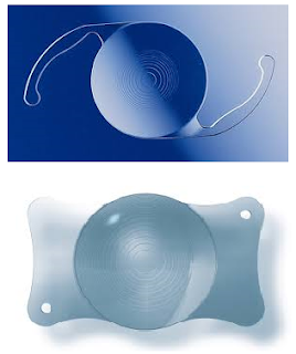
The Alcon Restor lens (above), and the AT LISA tri (below) are examples of bifocal and trifocal lens implants, respectively.


These diffractive multifocal lenses have rings etched onto either their front or back surfaces, which split/diffract incoming light rays into two foci. Some newer ones can even split the light into 3 foci. This fact gives rise to several implications.

1. Vision will be clear mainly at 2 (or sometimes 3) distances. For the older +4 Restor and the Tecnis multifocal, the near focus is at about 1 foot. So reading material has to be held quite close.

If it is held further away (say at 3 feet or so-intermediate distance), then it is not so clear anymore. 

Then things much farther away, say 15-20 feet away, start to become pretty clear again. (See the figure below-only the AT LISA tri is clear at intermediate distance)

Note: This is only the case if the surgeon has been very accurate in calculating the lens power/biometry.


Simulated clarity of images at various distances with different multifocal lens implants. The top 3 are bifocals, and the bottom one is a trifocal. Only the trifocal gives pretty good intermediate vision-but at some cost in terms of contrast when compared with the Alcon SV25T0 and AMO ZKB00. (From IOVS 2015;56:3970)

2. Because of the light splitting, there is a drop in contrast compared with a monofocal lens. Patients with multifocal lenses tend to be more sensitive to low light conditions-ie when it starts to get a little dark, they start to feel the drop in vision faster than patients with monofocal lenses. A person with a monofocal lens and reading glasses could read in dimmer conditions than a person with a multifocal lens.

3. The rings on the IOL give rings of light (halos) around light sources. This can be an issue when driving at night because of the halos around car headlamps. Having said this, many patients get used to the halos and do not notice it as much after a time.

Simulated halos around lights from a mutifocal lens

The above of course does not completely apply to the Crystalens or the Oculentis MPlus. The Crystalens is an accommodative lens, which gives somewhat unpredictable results-some patients don't get any near effect at all. 

The MPlus (available in Europe/Asia) has a wedged shaped section on the lens to focus for near. Point 1 above applies to this lens as well. However, although this lens does not give rings around lights, it can still cause 'halos' of a different shape-it is more like a smearing effect in one direction. Occasionally if a patient's pupil is small and not aligned with the lens, the patient may only have a far focus or a near focus and not both.

Last but not least, there is the Symfony lens which I covered in my last post. As mentioned, this is an 'in between' lens, ie in between a monofocal and a multifocal. It has no intermediate vision problems, and less halos than the usual multifocals, but does not give quite as good very near vision.

Currently, I offer patients a choice between a monofocal, an in between lens like the Symfony, and a full fledged trifocal like the AT LISA tri. I believe there is little to no benefit to be gained from using older bifocals such as the Restor and Tecnis Multifocal currently. Exactly which of those 3 choices gets implanted comes after a careful discussion with my patient regarding the pros and cons of each option. (So that we get big smiles all round afterwards!)

Tuesday, September 15, 2015

What type of lens implant should I choose for my cataract surgery?

Taking a quick look through a few eye care forums often turns this up as a commonly asked question, and one that garners quite a number of readers. This is not surprising, since over the years, more and more types of lenses with different pros and cons have become available. To recap, cataract surgery involves removing the hazy natural lens of the eye (called a cataract), and replacing it with a new lens implant.

Some of the currently available lenses: Tecnis Symfony (left), Acrysof  Toric monofocal (middle), and Zeiss Trifocal (right)


With cataract surgery nowadays, it is not so much the phacoemulsification technology or whether laser is used that affects the outcome. Rather, the outcome is determined by how accurately your doctor can calculate the necessary lens power, and what kind of lens is implanted into the eye. The available implants include 'monofocal' lenses (good vision at one distance), monofocal toric lenses (monofocal but with added feature to reduce astigmatism), and multifocal lenses (of which Extended Depth of Focus (EDOF) lenses can be considered a subset of - these provide clear vision at more than one distance).

Important questions to answer:

1. Do I value distance vision without glasses more, or near vision without glasses more?

2. Would I mind wearing glasses of any kind, including reading glasses?

3. Would monovision (one eye set for clear distance vision without glasses, the other for near) be a suitable option? Any prior experience with monovision or any chance of simulating it?

4. Would I mind intermittent visual disturbances such as halos around lights?

Simulated halo around a light, as seen through a multifocal lens


In some clinics, a questionnaire including these questions is filled in by patients prior to their surgery, to help in deciding on the type of lens for them. Of course, there is the final issue of cost, with multifocal and especially multifocal toric lenses commanding a premium.

Let's talk about some case scenarios:

1. You are very used to wearing glasses, and would actually like to wear them after surgery.
If you were shortsighted (myopic) before cataract surgery, and you habitually took your glasses off to read, then you would be good with monofocal lens implants but aiming to leave behind -1.50 to -2.00D of shortsightedness, in both eyes. Conversely, if you were hyperopic, I find that most patients like emmetropia (good distance vision without glasses), and then wear reading glasses for near visual work.

2. You had experience of monovision before, and liked it.
This could have been achieved naturally, or via glasses/contact lenses/LASIK. In this situation, it would be good to replicate it via cataract surgery.

3.You hate wearing glasses of any kind, and are concerned that monovision might not suit you or have tried it before and it does not suit you. 
In this case, you could consider the Extended Depth of Focus (EDOF) lenses, or the trifocal lenses. These lenses work quite predictably but also can cause some visual disturbances such as halos/rings around lights especially in the dark. Of these varieties, the EDOF lenses such as the Tecnis Symfony tend to cause the least disturbance ie least halos and least reduction in contrast.

If the benefit of seeing clearly at most distances with both eyes outweighs the possibility of some halos, then these lenses can be considered.

Generally, I would continue to avoid 'accommodating' lenses such as the Crystalens, and other multifocal designs such as the Oculentis Mplus range of lenses.

Hopefully, this post gives an idea of the thought processes that go into choosing a lens implant for cataract surgery. There is no one 'best lens' for everybody, and hopefully in the future there will be even better technology that can be applicable to more poeple with fewer side effects. Questions? Please feel free to post comments below.

Sunday, March 8, 2015

Cuts of the eye and first aid tips

Corneal lacerations-cuts to the eyeball

Mr L, a trainee carpenter, was trying to remove a nail from a piece of wood. He was thinking of getting off early that day, because of a date that evening to celebrate his girlfriend's birthday. As he levered the nail with the claw of the hammer, he suddenly felt it give and the nail flew off, straight into his eye.


The cornea is the clear window at the front of the eye. It is also the part of the eye closest to the object being looked at. 

Being constantly exposed to the environment, corneal injuries are a fairly common problem. They range from foreign bodies such as metallic particles from drilling or welding work, to abrasions/scratches, lacerations and chemical injuries.

Corneal lacerations occur when a sharp object cuts or pokes the cornea. They may be partial thickness, or they may go through the full thickness of the cornea and affect the inner parts of the eyeball.

Apart from direct damage to the eye, corneal lacerations are serious injuries which may also damage the eye by causing an infection, or impair vision because of corneal scarring after the eye heals.

Causes of lacerations

Common causes include flying objects, which may happen, for example with a nail as above or while cutting grass with an uncovered mower and while drilling and hammering. Broken glass from road traffic accidents are also a notorious cause of lacerations.

At the time of injury, a severe sharp pain will be felt as the cornea is one of the most sensitive parts of the body. There is almost always a clear history of a foreign object cutting or flying into the eye.
After the injury, vison is commonly blurred, especially if the laceration is deep and close to the centre of the cornea. There may be increased sensitivity to light, tearing, and it may be difficult to open the eye.

What the doctor will do to examine the eye

The doctor will usually instil some local anaesthetic eyedrops first to numb the eye and allow comfortable examination. Usually a careful examination on a slit lamp microscope then follows, with the doctor determining whether there are any remaining foreign bodies, and also very importantly, the depth of the laceration. If the cornea suffered a full thickness cut, fluid may leak from inside the eye, or internal parts of the eye such as the iris may protrude outwards.

A full thickness laceration has occurred in the centre of Mr L's eye. The nail penetrated the cornea, and also the lens of the eye, which has become a trauma induced cataract. Fine black nylon stitches were used to close the laceration.

Treatment of corneal lacerations

First aid for suspected corneal lacerations
  • ·         Do not attempt to remove any foreign bodies or what appear to be foreign bodies/dirt (protruding parts of the eye may look like foreign bodies or dirt)
  • ·         Do not wash or press on the eye
  • ·         If possible, obtain an eye shield or fashion one from a plastic cup. This is easily done by cutting the cup around the rim about 1-2cm from the bottom, so that the cup now only has a shallow rim. (see below)
  • ·         This can be taped so that the rim rests on the bony edges of the eyebrow and cheek.
  • ·         Send the patient straight to a specialist eye clinic or the emergency department.


Cuts of the cornea require different types of treatment, depending on how long or large they are, and even more importantly, whether the eye is penetrated.

If the cut is small and only affects a partial thickness of the cornea, it is treated like a corneal scratch or abrasion. Usually, this type of injury heals well with some antibiotic eyedrops to prevent infection, and the removal of any remaining foreign bodies. A bandage contact lens is sometimes used to protect a long shelving laceration.

For a corneal laceration that cuts through the full thickness of the cornea, the situation is more complicated. Usually, the first step, which is performed by the eye surgeon in an operating theatre, is to clean the wound and to stitch up the cornea so that there is no longer any fluid leakage from the eye. If there is internal tissue protruding from the eye, this is carefully repositioned into the eye when possible. Where there is more extensive damage such as lens damage causing a cataract, this may be dealt with at a second stage when tests are done to allow selection of the most suitable lens implant for the patient.

Mr L had his corneal laceration stitched up, followed by a cataract removal operation and lens implant. He now has good vision in that eye, however, due to the cataract surgery now has 'presbyopia' in that eye because the lens implant cannot autofocus like his original lens could before.

Prevention of corneal lacerations

·      These injuries can be prevented by wearing protective eyewear when engaging in activities such as grass cutting, hammering or drilling.


Corneal lacerations are potentially very serious injuries of the eye. Besides possible internal eye injuries, infections can also occur and lead to impaired vision. Do take care and use safety eyewear when engaging in hazardous activities, as the old adage ‘prevention is better than the cure’ certainly holds true here.

Tuesday, November 18, 2014

Femtosecond laser and cataract surgery-technical post

This post is going to be pretty technical, and for those who find it tough reading and just want to find out more about this new development in  cataract surgery, please email me (poryongming@yahoo.co.uk) or write in the comments section below.
A number of years ago, some laser companies decided to expand the capabilities of femtosecond laser LASIK flap cutters to make them cut at a deeper level. This is based on the premise that you still have to go in and remove the cataract after the laser cutting is done.

In cataract surgery, the cutting bits involve
1. Corneal cuts (both main incisions as well as relaxing incisions)
2. Capsule cuts
3. Nucleus cuts 
Normally/conventionally, the corneal cuts are made with a metal or diamond blade. The front capsule opening is torn manually in a circular fashion by the surgeon. Finally the nucleus is cut (with ultrasound) or chopped (manually).
The femtosecond laser is a good cutting machine, however, cataract surgery also demands removal of the hazy lens material besides cutting, and the removal is still done with a conventional phacoemulsification machine.

The LenSx machine from Alcon

The Catalys machine from AMO
The Lensar machine
The Victus machine from Bausch and Lomb
The above are the currently available platforms for performing laser assisted cataract surgery. Each have their own pros and cons, and it may be worth asking your surgeon about the system he/she is using. 

Is there a difference in result between conventional vs laser cataract surgery? 

I think it is fair to say that any laser can make more uniform, consistent cuts than those made manually. However, there are several lines of evidence which suggest that there is no difference in clinical outcomes between laser assisted surgery or conventional phacoemulsification.

Effect of femtosecond laser-created clear corneal incision on corne... - PubMed - NCBI
J Cataract Refract Surg. 2014 Apr;40(4):531-7. doi: 10.1016/j.jcrs.2013.11.027. Epub 2014 Feb 20. Comparative Study; Randomized Controlled Trial; Research Support, Non-U.S. Gov't

Preview by Yahoo


Evaluation of femtosecond laser-assisted and manual clear corneal i... - PubMed - NCBI
J Refract Surg. 2014 Aug;30(8):522-5. doi: 10.3928/1081597X-20140711-04.

Preview by Yahoo


The above two papers show that there are no or insignificant differences between pre and post operative central corneal curvature, which influences surgically induced astigmatism and post-operative refractive predictability. One paper showed differences in the corneal topography immediately overlying the incision, but this is far in the corneal periphery (not affecting vision) and would be expected to settle down with time.

Effect of reducing ultrasound energy
The main aim of reducing ultrasound energy is to reduce corneal endothelial damage, rather than retinal problems. The phaco probe is normally too far from the retina to cause any problems with the ultrasound. It is, however, only millimeters away from the corneal endothelium during surgery.

Even in this regard, the data is mixed. One paper in fact reported greater early endothelial cell loss in laser cataract surgery eyes than conventional phacoemulsification. The other paper showed no significant differences.


Effect of femtosecond laser-assisted cataract surgery on the cornea... - PubMed - NCBI
J Cataract Refract Surg. 2014 Sep 9. pii: S0886-3350(14)01099-2. doi: 10.1016/j.jcrs.2014.05.031. [Epub ahead of print]

Preview by Yahoo


Endothelial cell loss and refractive predictability in femtosecond ... - PubMed - NCBI
Acta Ophthalmol. 2014 Nov;92(7):617-22. doi: 10.1111/aos.12406. Epub 2014 Jun 2.

Preview by Yahoo


Effect on IOL position and refractive accuracy
One of the papers below suggests better IOL position after laser cataract surgery, and the other says there is better refractive predictability. Interestingly, the difference in post operative mean refractive error is miniscule: 0.12D. And they say the difference was greater in very short or very long eyes, suggesting a problem with the formula they were using rather than the surgery itself.

Comparison of IOL power calculation and refractive outcome after la... - PubMed - NCBI
J Refract Surg. 2012 Aug;28(8):540-4. doi: 10.3928/1081597X-20120703-04. Epub 2012 Jul 13. Comparative Study

Preview by Yahoo


There is no study which compares relatively minor problems like conjunctival haemorrhage etc between laser and conventional surgery that I am aware of. Part of the reason I think is that we just don't see some of these things with conventional surgery. Journal editors tend not to want to publish things that are already known...

With regard to one of the other problems cited with femtosecond laser cataract surgery-that of intraoperative miosis (pupil getting smaller), it is something that does not happen in conventional phacoemulsification unless there is IFIS (Intraoperative Floppy Iris Syndrome), related to the use of alpha antagonists for prostate problems. Nowadays, this problem is minimised by doing the lens removal as soon as possible after the laser cutting procedure.
Is femtosecond laser assisted cataract surgery better, or gives better outcomes than, conventional phacoemulsification cataract surgery? I think the jury is still out on this one!

Saturday, November 1, 2014

Cataract and Cataract Surgery FAQs

In my line of work, I get asked lots of questions about cataracts, quite simply because they are one of the most common causes of blurred vision as one gets older.

Here goes:

1. What are cataracts? Are they a growth in the eye?

Cataracts are not growths.  One of the parts of the eye, called the lens, undergoes ageing processes over the years. A lens, like a camera or spectacle lens, needs to transmit and focus light and therefore needs to be transparent. Unfortunately age causes many lenses to become hazy and this blocks vision, much like trying to look through a dirty window.

2. How do I know if I have cataracts?

The main problem with cataracts is blurred vision. Of course, there are many other causes of blurred vision, such as shortsightedness or astigmatism. Therefore, if vision is blurry, try wearing spectacles first and see if the vision is cleared up.

If so, the problem is probably just spectacle power, or perhaps the cataracts are very mild. Significant cataracts will cause blurred vision even if spectacles are worn. Generally speaking, if vision cannot be made clear with glasses, one should visit the eye doctor to make sure there are no serious eye problems.

3. How does it feel like to have cataracts?

This is related to the prior question. Although we may think that cataracts are just a hazy lens and are the same, in fact there are a few types of cataracts that affect vision in slightly different ways. For example, vision can be clearer under certain conditions, or sometimes patients get double or triple vision.

A yellow nuclear sclerotic cataract
One type of cataract causes the lens to become yellowish (above). This is called 'nuclear sclerosis', and the eye also becomes more and more shortsighted. Patients with this type of cataract may need to increase their shortsighted spectacle power several times a year. On the other hand, long sighted people may find their spectacle power getting less and less, and near vision getting better and better! This is sometimes called second sight.

Another type of cataract causes glare. This means that a person can see reasonably well in the dark, but when there is a bright light shining from in front, vision undergoes a 'white out', ie everything turns bright white and vision suddenly becomes very blurry. This happens with a cataract called a 'posterior subcapsular' cataract.

A very dense, white cataract
If cataracts are neglected for a long time, they can turn white (regardless of which type they originally were). White cataracts are more dangerous than other types, because they sometimes absorb water and swell. A swollen lens can physically block the drainage angle of the eye, or leak proteins which also block the drainage angle. This causes a sharp spike of eye pressure, leading to a type of glaucoma and eye pain. This is the only situation where cataracts are associated with eye pain.

4. What causes cataracts?

As mentioned, most cataracts are due to age related changes in the eye. Proteins break down, clump together, sometimes water is even absorbed into the lens causing to swell.

Besides age, there are other less common causes of cataracts. Certain medicines such as steroid eyedrops or tablets, if taken in high doses for too long a time, can cause cataracts to develop. Diabetes, which results in too much sugar in the blood, also cause cataracts to develop earlier, especially if the blood sugar levels are not controlled properly and become very high for a lot of the time. Cigarette smoking is well known to be associated with earlier cataract development as well. Finally, bad injuries to the eye, such as a direct punch or a hit in the eye can also cause a cataract to develop.

5. How can I prevent cataracts, or can I slow down their progression?

Firstly, any risk factors such as diabetes or cigarette smoking should be addressed. Modifying these risk factors will have the biggest impact on slowing cataract progression.

Secondly, eat a healthy diet full of antioxidants. In general, it is better to get these naturally from green leafy vegetables and colourful fruits, rather than supplements. However, certain nutrients may only be obtainable in large quantities in supplements.

Studies of big groups of people suggest that eating more fruits and vegetables can delay cataract development, but nothing conclusive so far can be said for individual nutrient supplements such as vitamin C.

6. How are cataracts treated?

Cataracts are treated with surgery to remove them, and at the same operation they are replaced by lens implants made of a special acrylic material.


Cataract surgery is carried out under local anaesthesia, and is painless. The hazy lens material is broken up with ultrasound, and then removed through a very small incision about 2mm long. The lens implant can then be folded and injected into the eye through this very small opening.

Recovery is very quick, with most patients seeing very well even on the day after surgery.

Sometimes patients ask me if they should wait until after their holiday to have surgery. I tell them that if time permits, to do it before, as they will enjoy the view so much more!

7. What are the latest advances in cataract surgery?

The most important developments in cataract surgery have to do with how spectacles can be avoided after surgery. Yes, no matter how high the spectacle power before surgery, most of the time spectacle wear can be avoided after surgery. How is that possible?

Focusing of light in the eye depends on the cornea, the lens, and how long the eyeball is. The shape of the cornea can be changed (in LASIK), and the lens power can be changed (in cataract surgery) to reduce or eliminate spectacle power.

A normal spherical Tecnis (AMO) lens
A Toric Tecnis (AMO) lens. Note the dots arranged in a line at the sides of the lens. This allow the surgeon to place the lens in exactly the right position to reduce or eliminate astigmatism after surgery
The Tecnis Symfony (AMO) lens. This is a multifocal lens that is also available in a toric version (right) so it can be used in most patients

Nowadays, lens implants also come in 'toric' versions to correct astigmatism, and there are also 'multifocal' lenses that give good near and far vision to avoid reading glasses after cataract surgery. 

For patients who would like clear far and near vision without glasses, and have astigmatism, there are even multifocal+toric lens implants.

Multifocal lens implants can give halos around lights at night, however, this has been minimised with newer extended depth of focus (EDOF) implants like the Tecnis Symfony (above).

Laser cataract surgery

Some surgeons are now using a type of laser to help to do certain steps of the surgery. They call this bladeless cataract surgery, much like how LASIK became 'bladeless' LASIK.


Unfortunately, using this laser to help with surgery makes the surgery much more expensive and also can result in problems that probably would not have happened if the surgery had been done with conventional phacoemulsification surgery. In the article above, 1/3 of patients had conjunctival haemorrhage, and 1/5 had anterior capsular tags. In 1/3 of patients, the pupil became smaller, making the surgery more difficult for the surgeon and increasing the risk of other complications.

Although this technology may become better with time, and surgeons will become more experienced with using it, currently this is an example of how a 'latest advance' is not necessarily better and in fact has become a kind of marketing tool.

Below is an exchange in the local newspapers in 2013, where the Singapore Academy of Medicine published a response in the Straits Times to counter certain unfounded claims put forth by proponents of the laser cataract surgery procedure:
http://ams.edu.sg/view-pdf.aspx?file=media%5C499_fi_807.pdf&ofile=STForum20130516+-+Traditional+cataract+surgery+remains+%27gold+standard%27.pdf